Leg Bone Diagram ~ Hip Joint Anatomy Hip Bones Ligaments Muscles. Pelvic bone labeled 12 photos of the pelvic bone labeled pelvic bone labeled, pelvic bone labeling quiz. The lower leg contains two major long bones, the tibia and the fibula, which are both very strong skeletal structures. Most leg pain results from wear and tear, overuse, or injuries in joints or bones or in muscles, ligaments, tendons or other soft tissues. For diagram showing its location relative to the fibula, tibia, patella, and other bones of the leg. The major bones of the leg are the femur (thigh bone), tibia (shin bone), and adjacent fibula, and these are all long bones.the patella (kneecap) is the sesamoid bone in front of the knee.most of the leg skeleton has bony prominences and margins that can be palpated and some serve as anatomical landmarks that define the extent of the leg.
Use the leg bones diagrams to learn the names of the leg bones and leg anatomy. Its lower end helps create the knee joint. The pubis, ischium, and ilium together constitute the pelvis while the thigh bone is the femur. The humerus and the femur are corresponding bones of the arms and legs, respectively. The structure of a long bone allows for the best visualization of all of the parts of a bone ().a long bone has two parts:
Pin By Genna Hornsby On Anatomy Human Anatomy And Physiology Medical Anatomy Anatomy Bones from i.pinimg.com Related posts of muscles and tendons of the leg muscle anatomy in shoulder. At the same time, the bones and joints of the leg and foot must be strong enough to support the body's weight while remaining. The major bones of the leg are the femur (thigh bone), tibia (shin bone), and adjacent fibula, and these are all long bones.the patella (kneecap) is the sesamoid bone in front of the knee.most of the leg skeleton has bony prominences and margins that can be palpated and some serve as anatomical landmarks that define the extent of the leg. These muscles work together to produce movements such as standing, walking, running, and jumping. The humerus and the femur are corresponding bones of the arms and legs, respectively. This area is commonly referred to as the calf. The tibia and fibula are two long bones that run parallel to each other, forming the scaffold of the leg and providing attachment points for many muscles. The tibia (also called the shinbone) is located near the midline of the leg.
The tibia, commonly known as the 'shin bone', is the largest and most medial of the two.you can palpate its anterior border when you run your finger down the anterior aspect of your leg.
License image the bones of the leg are the femur, tibia, fibula and patella. 2006 kia optima belt diagram. Related posts of diagram of leg bones pelvic bone labeled. Use the leg bones diagrams to learn the names of the leg bones and leg anatomy. See more ideas about muscle anatomy, leg muscles anatomy, anatomy. The thigh bone, or femur, is the large upper leg bone that connects the lower leg bones (knee joint) to the pelvic bone (hip joint). For diagram showing its location relative to the fibula, tibia, patella, and other bones of the leg. The lower leg contains two major long bones, the tibia and the fibula, which are both very strong skeletal structures. Browse 7,052 leg bone stock photos and images available, or search for leg bone xray or human leg bone to find more great stock photos and pictures. Most leg pain results from wear and tear, overuse, or injuries in joints or bones or in muscles, ligaments, tendons or other soft tissues. The knee joint is the largest joint in the body and is primarily a hinge joint, although some sliding and rotation occur. Leg bone anatomy diagram diagram of human leg human anatomy diagram 10 / 10 ( 1 vote ) in this image, you will find femur, medial epicondyle of the femur, patella, tibial tuberosity, anterior rest of the tibia, a medial surface of the tibia, lateral epicondyle of the femur, head of the fibula, fibula, medial malleolus of the tibia, lateral. Its lower end helps create the knee joint.
The bones of the leg are the femur, tibia, fibula and patella.the foot bones shown in this diagram are the talus, navicular, cuneiform, cuboid, metatarsals and calcaneus. Infographic diagram of human skeleton lower limb anatomy bone system or leg bone anterior view lower limb bones (thigh, leg and foot) labeling page clinical anatomy: Most leg pain results from wear and tear, overuse, or injuries in joints or bones or in muscles, ligaments, tendons or other soft tissues. The tibia, commonly known as the 'shin bone', is the largest and most medial of the two.you can palpate its anterior border when you run your finger down the anterior aspect of your leg. The pubis, ischium, and ilium together constitute the pelvis while the thigh bone is the femur.
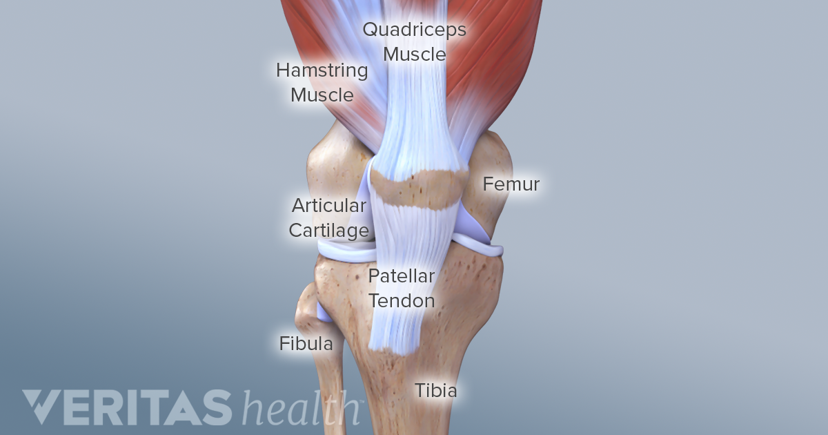
Knee Anatomy from embed.widencdn.net Its lower end helps create the knee joint. The humerus and the femur are corresponding bones of the arms and legs, respectively. The medial, larger bone of the lower leg. The major bones of the leg are the femur (thigh bone), tibia (shin bone), and adjacent fibula, and these are all long bones.the patella (kneecap) is the sesamoid bone in front of the knee.most of the leg skeleton has bony prominences and margins that can be palpated and some serve as anatomical landmarks that define the extent of the leg. The diaphysis and the epiphysis.the diaphysis is the tubular shaft that runs between the proximal and distal ends of the bone. Muscle system diagram photos muscular system more than 133 muscle chart back at on the anterior and posterior views. The femur, or thighbone, is the longest and largest bone in the human body. The majority of muscles in the leg are considered long muscles, in that they stretch great distances.
The femur, or thigh bone, is the single bone of the thigh region (figure 6.51).
The knee joint is the largest joint in the body and is primarily a hinge joint, although some sliding and rotation occur. Upper leg bones diagram the junction of where these structures converge at the pubic bone revolves around the inguinal canal bodies and the intervening discs from the lower. The bones of the leg and foot form part of the appendicular skeleton that supports the many muscles of the lower limbs. The major bones of the leg are the femur (thigh bone), tibia (shin bone), and adjacent fibula, and these are all long bones.the patella (kneecap) is the sesamoid bone in front of the knee.most of the leg skeleton has bony prominences and margins that can be palpated and some serve as anatomical landmarks that define the extent of the leg. These landmarks are the anterior superior iliac spine. Learn vocabulary, terms, and more with flashcards, games, and other study tools. 2006 kia optima belt diagram. The structure of a long bone allows for the best visualization of all of the parts of a bone ().a long bone has two parts: The hip itself is a ball and socket joint, much like the shoulder.the structures necessary to create this joint are the socket, the joint capsule, muscle, ligaments, and the neck. Related posts of diagram of leg bones pelvic bone labeled. The femur, or thighbone, is the longest and largest bone in the human body.it is the only bone in the upper leg. The humerus and the femur are corresponding bones of the arms and legs, respectively. Educational diagram with pronated, normal and supinated compared examples with bone titles.
Muscle anatomy in shoulder 12 photos of the muscle anatomy in shoulder muscle anatomy neck and shoulder, muscle anatomy of shoulder, muscle anatomy of shoulder joint, muscle anatomy shoulder back, muscle anatomy shoulder upper arm, human muscles, muscle anatomy neck and shoulder, muscle anatomy of shoulder, muscle. The bones of the knee and leg dummies The major bones of the leg are the femur (thigh bone), tibia (shin bone), and adjacent fibula, and these are all long bones.the patella (kneecap) is the sesamoid bone in front of the knee.most of the leg skeleton has bony prominences and margins that can be palpated and some serve as anatomical landmarks that define the extent of the leg. Its lower end helps create the knee joint. The bones of the leg and foot form part of the appendicular skeleton that supports the many muscles of the lower limbs.
6 3 Bone Structure Anatomy Physiology from open.oregonstate.education License image the bones of the leg are the femur, tibia, fibula and patella. At the same time, the bones and joints of the leg and foot must be strong enough to support the body's weight while remaining. The tibia, commonly known as the 'shin bone', is the largest and most medial of the two.you can palpate its anterior border when you run your finger down the anterior aspect of your leg. Muscle anatomy in shoulder 12 photos of the muscle anatomy in shoulder muscle anatomy neck and shoulder, muscle anatomy of shoulder, muscle anatomy of shoulder joint, muscle anatomy shoulder back, muscle anatomy shoulder upper arm, human muscles, muscle anatomy neck and shoulder, muscle anatomy of shoulder, muscle. These muscles work together to produce movements such as standing, walking, running, and jumping. The head of the fibula. The medial, larger bone of the lower leg. As these muscles contract and relax, they move skeletal bones to create movement of the body.
These muscles work together to produce movements such as standing, walking, running, and jumping.
Most leg pain results from wear and tear, overuse, or injuries in joints or bones or in muscles, ligaments, tendons or other soft tissues. The bones of the leg are the femur, tibia, fibula and patella.the foot bones shown in this diagram are the talus, navicular, cuneiform, cuboid, metatarsals and calcaneus. The bones together make up the hip. The lower leg extends from the knee to the ankle. The bones of the leg and foot form part of the appendicular skeleton that supports the many muscles of the lower limbs. Browse 7,052 leg bone stock photos and images available, or search for leg bone xray or human leg bone to find more great stock photos and pictures. The tibia (also called the shinbone) is located near the midline of the leg. These landmarks are the anterior superior iliac spine. The femur, or thighbone, is the longest and largest bone in the human body. See more ideas about muscle anatomy, leg muscles anatomy, anatomy. The diaphysis and the epiphysis.the diaphysis is the tubular shaft that runs between the proximal and distal ends of the bone. Learn vocabulary, terms, and more with flashcards, games, and other study tools. This area is commonly referred to as the calf.
Share :
Post a Comment
for "Leg Bone Diagram ~ Hip Joint Anatomy Hip Bones Ligaments Muscles"


Post a Comment for "Leg Bone Diagram ~ Hip Joint Anatomy Hip Bones Ligaments Muscles"